Deformity of the femur. Varus deformity of the lower extremities in children: causes, photos, treatment Varus deformity of the hips
Predominantly valgus deformity of the hip joints in children is diagnosed during a routine medical examination by an orthopedist. The pathological condition is quite rare. Both boys and girls are equally affected. A number of factors can provoke an ailment, which are divided into congenital and acquired. If you do not carry out timely therapy of the disease, serious complications appear.
Why does a pathological condition develop?
Contributes to the appearance of valgus deformity of the hip joints in young patients with a partial lesion in the lateral part of the epiphyseal cartilage located above the head of the bone. Pathology often develops during life in children due to untreated joint dysplasia. During childbirth in infants, the femoral head is located in physiological valgus and turned back. In the process of growing up, the ratio changes. In adults, the cervical-diaphyseal angle is predominantly 120°. The anteversion angle is approximately 10°. If violations are observed, then in small patients these angles change, due to which valgus deformity of the hip joints develops. In addition, the following factors influence the development of this pathological condition:
- cerebral palsy;
- transferred poliomyelitis;
- dystrophy of muscle tissue;
- exostosis;
- cancerous diseases.
In addition, in exceptional situations, hallux valgus is provoked by a displaced fracture of the femoral neck and rickets.
What symptoms are observed?
 If the pathology develops on one side, then the child develops lameness.
 Mostly, when a child is diagnosed with bilateral damage to the hip joints, the pathology does not manifest itself in any way. If a unilateral violation is observed, then most often the limb on this side lengthens, as a result of which the gait changes, the small patient begins to limp on one leg. The pathological condition is difficult to detect, as the function of the hip joint is preserved.
With the help of X-ray examination, it is possible to detect the disease when the child is one year old. In this case, the femur is bent and creates a right angle. The epiphyseal cartilage is localized almost vertically, and the head of the bone can be enlarged, but it is located in a vertical cavity. If the cervical-diaphyseal angle is less than 110°, then the depression is flat and shallow. If it reaches 130°, the depression develops in the usual way. The trochanter is located above the neck and has a medial slope. In the process of development of hallux valgus, it increases.
Diagnosis of valgus deformity of the hips in children
When parents suspect that a child is developing valgus deformity of the femoral neck, it is important to immediately contact a medical facility. First of all, the orthopedist conducts a visual examination. Then the little patient is sent for an X-ray examination, during which an internal rotation of the limb is necessarily made. In addition, ultrasound examination of the joints and computed or magnetic resonance imaging are sometimes required.
How is the treatment carried out?
 Orthopedic shoes will help to cope with the problem.
 Conservative therapy, which involves stretching or immobilizing the hip joint, is ineffective for hallux valgus. They resort to the help of the only method of conservative treatment, which is to wear. With the help of it, it is possible to prevent the appearance of deformation of the distal legs. Thanks to the insole, which is put into the shoes, it turns out to even out the length of the lower extremities and compensate for the shortening of the affected leg.
Surgery in the fight against a pathological condition
Therefore, patients suffering from valgus deformity of the hip joint are prescribed surgery. Its appearance is directly related to the magnitude of the deformation, the severity of the course of the disease and the age category of the patient. If the angle of curvature of the hip is 50 °, then the operation is not prescribed. It is enough to constantly monitor the patient and conduct an X-ray examination every 6 months. Surgical intervention with such a curvature is resorted to in situations where the deformation is actively progressing.
Direct indications for surgery are:
- an increase in the angle of more than 60 ° degrees;
- decreased strength of the gluteus medius;
- severe impairment of walking.
A contraindication is the absence of unwanted symptoms and progression if the angle of curvature is 60 degrees.
 Operationally, you can completely eliminate the defect.
 As for the age of the patient, the operation is rarely performed on children who are not yet 2 years old. This is due to the fact that during this period the deformation of the femurs and joints is not observed very clearly. However, surgery at an earlier age has a significant advantage, which is the ability to completely remodel the affected bone tissue. Thanks to the operation, it is possible to get rid of the valgus curvature of the hip, to reduce the shift of the joint. In addition, after the operation, the length of the legs is leveled.
Ticket 36:
1 ) Fractures of the condyles of the femur and tibia: classification, diagnosis, treatment. There are fractures of one of the condyles and both condyles of the femur (intercondylar Y- and T-shaped). Isolated fractures of the condyles usually occur with a sharp deviation of the lower leg inwards (fracture of the internal condyle) or outwards (fracture of the external condyle). Fractures of both condyles often occur as a result of a fall from a great height onto a straight leg. Clinic. With an isolated fracture of the external condyle with displacement of fragments, a valgus deviation of the lower leg (genu valgum) occurs, with a fracture of the internal condyle with displacement, a varus deviation of the lower leg (genu varum). With fractures of both condyles with displacement, anatomical shortening of the limb can be detected. In addition, the joint is sharply enlarged in volume due to hemarthrosis, the limb takes a forced position: the leg is slightly bent at the knee and hip joints. Active and passive movements in the knee joint are sharply painful. On palpation, there is an increase in pain and a symptom of balloting of the patella. The following clinical symptoms are characteristic of fractures of this localization: Pain in the knee joint and the lower part of the thigh, aggravated by palpation and pressure on the condyles. varus or hallux valgus knee joint. Thigh circumference in the region of the condyles increased.contours knee joint smoothed out.Fluctuation at the knee ( hemarthrosis).Ballotion of the patella. Passive movements in the knee joint possible but painful.Sometimes can be defined bone crunch.The diagnosis is clarified by radiographs made in two projections. Treatment. Fractures of the distal femur without displacement of fragments are treated by immobilization with a plaster cast (3-5 weeks) or by I. R. Voronovich: lateral compression osteosynthesis with pins with thrust pads is used. This method allows you to perform all 4 principles of treatment of intra-articular injuries: Ideal reposition fracture (with an accuracy of 2 mm, since only with such a displacement of the articular surfaces is it possible to regenerate hyaline cartilage). Reliable fixation fragments for the entire period of consolidation. Early feature(for the full function of cartilage and its metabolic processes). on the damaged joint. Before fixation, puncture of the knee joint for the purpose of evacuating blood and introducing into the joint 20-30 ml of 1% novocaine solution. During the first 7-10 days after injury, it often becomes necessary to repeat punctures of the joint and evacuate blood, which is one of the ways to prevent post-traumatic arthrosis. traction. The load at a fracture without displacement is 2-4 kg, with displacement - 4-8 kg. The traction period is b weeks, the leg is fixed with the son-in-law Circular plaster cast to the groin for a period of 6 weeks. After removing the bandage, they begin restorative treatment: baths, paraffin, massage, exercise therapy, mechanotherapy. Recovery disability for fractures without displacement of fragments after 3-3.5 months; with displacement of fragments - after 5-6 months. Surgical treatment: shown when bone fragments are not juxtaposed in a closed way. Bone fragments are exposed, repositioned and fixed either with a plate or with 1-2 metal rods. The operated leg is fixed with a plaster cast until a callus is formed. Then proceed to rehabilitation treatment. Surgical intervention allows more accurate repositioning of fragments, their strong fixation and, due to this, earlier start of functional treatment (2-3 weeks from the moment of surgery). Full load on the injured limb is allowed no earlier than after 3.5-4.5 months. Fractures of the condyles of the tibia. Fractures of the condyles of the tibia are intra-articular injuries and occur most often when falling on straight legs or when the lower leg deviates outward or inward. There are fractures of the external condyle, internal condyle, as well as T- and Y-shaped fractures of both condyles. Fractures of the condyles can be impression and chipping type. They may be accompanied by damage to the meniscus, the ligamentous apparatus of the knee joint, fractures of the intercondylar eminence of the tibia, fractures of the head of the fibula, etc. Clinical picture in fractures of the condyles of the tibia, it corresponds to intra-articular damage: the joint is enlarged in volume, the leg is slightly bent, hemarthrosis is detected by the symptom of balloting the patella. The tibia is deflected outwards in case of a fracture of the external condyle or inwardly in case of a fracture of the internal condyle. The transverse size of the tibia in the area of the condyles is increased in comparison with the healthy leg, especially in T- and Y-shaped fractures. On palpation, the area of the fracture is sharply painful. Characterized by lateral mobility in the knee joint with unbent lower leg. There are no active movements in the joint, passive movements cause sharp pain. The patient cannot raise the straightened leg. Sometimes damage to the external condyle is accompanied by a fracture of the head or neck of the fibula. In this case, the peroneal nerve can be damaged, which is recognized by a violation of sensitivity, as well as motor disorders of the foot. X-ray examination allows you to clarify the diagnosis and identify the features of the fracture. Treatment. In case of fractures of the condyles of the lower leg without displacement, a joint is punctured for aspiration of blood and the introduction of 20-40 ml of a 1% solution of novocaine. The injured limb is fixed with a circular plaster cast. From the 2nd day, exercises for the quadriceps femoris are recommended. Walking with crutches without weight on the affected leg is allowed after a week. The plaster bandage is removed after 6 weeks. Loading of the leg is allowed 4-4.5 months after the fracture. With early loading, impression of the damaged condyle may occur. In case of a displaced condyle fracture, both conservative and surgical treatment is used. In some cases, fractures with displacement, especially comminuted, T and V-shaped fractures, can be applied permanent skeletal traction. At the same time, the patient's limb is placed on the Beler splint, the needle is passed through the calcaneus, the load along the axis of the lower leg is 4-5 kg. The duration of treatment with this method is 4-5 weeks, after which the limb is fixed with a gypsum bandage. Further treatment is the same as for a fracture of the condyles without displacement of the fragments. A physiological method with good results of treatment was proposed by I. R. Voronovich. Surgical treatment is indicated for unsuccessful conservative treatment. The operation is performed 4-5 days after the injury: open reposition of the fracture and osteosynthesis with metal structures. The sutures are removed on the 12-14th day, and further management of the patient, as in case of fractures of the condyles without displacement.
2) .Conservative treatment of osteoarthritis of large joints. Methods of the department. Patients with deforming arthrosis it is necessary to observe a certain motor mode aimed at unloading the diseased joint. They should avoid walking for a long time, standing on their feet for a long time or staying in one position, and should not carry weights. In case of severe pain while walking, it is necessary to use a cane or walk with crutches. To unload a diseased joint, even at home, cuff traction with a load along the leg axis of 2-3 kg should be used. With sharp pains that do not go away from the above measures, you can apply fixation of the joint with a plaster cast for 2-4 weeks, but at the same time movements are even more limited, and contractures are aggravated. The goal of conservative treatment of arthrosis– restoration of blood circulation in the tissues of the diseased joint. Therapy should be comprehensive and include not only drug treatment, but also physiotherapy, spa treatment. The conservative treatment described below should be comprehensive and correspond to the stage of development of the disease. Means of microcirculatory influence used to restore the microcirculation system. For this purpose, various agents are used, the pharmacogenesis of which is not the same: angiotrophin, andekalin, depokallikrein, dilminal, inkrepan. They are prescribed in the first stage of the disease in patients without synovitis within 3 weeks. With the development of inflammation in the tissues of the joint, it is better to use agents that inactivate the kinin system - countercal, zalol, trasilol, etc.
One of the rare anomalies in the development of the femur is their deformity of the varus type. The disease occurs no more often than in 0.3-0.8% of cases among newborns. Along with valgus curvature of the proximal end of the femur, congenital varus deformity of the femur is a skeletal defect. It can lead to serious violations of the functioning of the musculoskeletal system.
Description of varus deformity of the hips
The basis of the curvature of the hip joints according to the varus type is the shortening of the neck of the femur and a decrease in the degree of the neck-diaphyseal angle. Another name for the disease is juvenile epiphysiolysis, although in fact the latter is one of the forms of hip deformity and is very rare, only in adolescence. Changes in the hip joints in this pathology are significant - dystrophy of the spongy tissue of the neck, bone destruction, formation of cysts, fibrosis phenomena.
Varus-type hip deformity includes a whole range of symptoms from the lower extremities. With this disease, the following symptoms can be combined:
- change in the shape of the articular heads of the pelvic bones;
- leg shortening;
- contractures of the hip joint;
- dysplasia, dystopia of leg muscles;
- lumbar lordosis.
In a child with hip deformity, a serious violation of rotation and abduction of the leg is noted, so the gait becomes “duck”. When an anomaly occurs in a newborn, then the leg is already shorter than the second one from birth, and the trochanter of the thigh is located higher than expected. If the pathology is not treated for a long time, it continues to progress, ossification occurs. The femoral neck is bent, the diaphysis is shortened.
When the deformity of the joint according to the varus type occurs at the age of 3-5 years, a trihedral bone fragment appears in the femoral neck. Visually, the head and neck of the femur resemble an inverted U. The joint space becomes tortuous, the edges of the bone are jagged, uneven, and foci of sclerosis appear in them. After the gap of the hip joint expands to 1-1.2 cm, the neck is shortened, and the head ceases to develop normally.
If in the childhood form of varus curvature there are changes in the bone zone, then in the youthful form there are violations in the growth zone. The latter is loosened, the bone is resorbed, the head slowly slides down. Therefore, the pathology is called "epiphyseolysis of the femoral head."

Causes of the disease
Usually, varus deformity of the congenital type becomes a consequence of intrauterine damage to the cartilage of the femur or a violation of the process of their compaction. In 2/3 of the patients the defect is unilateral, in other cases it is bilateral. Thus, the disease occurs due to various problems in the embryonic period, which can happen for such reasons:
- severe maternal infections during pregnancy;
- alcohol abuse, drug use, toxic drugs;
- poisoning, intoxication;
- advanced age of the mother;
- endocrine diseases;
- the influence of radiation.
As for the acquired forms, they can occur for various reasons. So, juvenile epiphysiolysis develops at 11-16 years of age - during puberty, or precedes puberty. The head of the femur begins to deform precisely against the background of a general restructuring of the body, when some parts of the skeleton are most vulnerable. It is believed that hormonal disorders become the cause of pathology in adolescents, therefore other phenomena are also often noted in patients:
- lack of secondary sexual characteristics;
- delay in menstruation in girls;
- too tall;
- obesity.
Also, varus deformity of the femur is associated with trauma and rickets, with a number of systemic diseases - pathological bone fragility, fibrous osteodysplasia, dyschondroplasia. Also, the cause of the pathology can be:
- failed hip surgery
- osteomyelitis;
- bone tuberculosis;
- osteochondropathy.

Symptoms of the disease
The children's form of pathology usually begins to develop no later than 3-5 years, since during this period there is an increase in the load on the lower limbs. Parents may experience the following symptoms:
- lameness after a long walk;
- pain in the legs in the afternoon due to fatigue;
- inability to run for a long time, play outdoor games;
- discomfort in the thigh and knee, in the popliteal region;
- pain in the knee joint.
Over time, the leg on the affected side becomes somewhat thinner, hip abduction becomes more difficult, and more so on the inside (on the contrary, it can increase on the outside). In a number of children, the symptomatology begins with pain in the knee, and it is not always possible to establish a connection with a hip lesion immediately.

Juvenile forms of hip deformity often do not give clinical signs at all, only in advanced cases they begin to manifest themselves. There is a slight lameness, increased fatigue when walking. Some adolescents have a protruding stomach, there is a pathological lordosis of the spine. The strength and tone of the gluteal muscles are reduced. With bilateral damage to the hips, the child begins to walk like a duck, waddling, swaying.
Classification of pathology
Due to the appearance and radiological signs, the deformity of the femur can exist in the following forms: children's, youthful, symptomatic, rickets, tuberculosis. Also, the disease is isolated or is reflected in other joints, feet of the child. Another classification involves the division of the disease into three stages (degrees).
First degree of severity
At the first stage, pathological changes begin in the growth region of the femur. Gradually, its loosening and expansion occurs, but the epiphysis does not move.
Second degree of severity
At the second stage, progression of the processes of bone tissue restructuring and displacement of the epiphysis is observed. The picture shows a thinning of the femoral neck, the fuzziness of its structure.
Third degree of severity
At the third stage, a complication of the pathology is already registered - deforming arthrosis. There is also atrophy of the muscles of the lower limb and marked changes in gait.

Diagnosis of varus deformity
The most popular and informative method of diagnosis is radiography of the hip joint. At the very beginning of the hip deformity, heterogeneity of the bone mass in the region of the femoral neck is revealed. Later, other structural changes appear, as well as violations of the anatomy of the hip joint.
When examined by an orthopedist, violations in adduction and abduction of the limb on one or both sides are revealed. In parallel, kyphosis, scoliosis, lordosis, wedge-shaped deformity of the vertebrae, and other disorders in the knees, sacrum, and ankles can be diagnosed.
Treatment of pathology
In the early stages, conservative therapy helps well, later surgical treatment is used. Initially, as a rule, the patient is hospitalized for limb traction (skeletal traction), after which treatment is continued at home.
Conservative treatment
With a congenital form of pathology, conservative therapy is indicated for all babies up to 3 months of age. The goals are to normalize the blood supply to the hip joint and accelerate bone recovery, improve the tone of muscle tissue, and reduce the effect of muscles on the condition of the joint. For this, the following therapies are performed:
- wide swaddling for 14 days, after - Freyka pillow for 2.5 months;
- sollux, paraffin applications;
- from 6-8 weeks - electrophoresis on the joint area with calcium, phosphorus, with vasodilators - on the area of the sacral spine.

Other forms of varus deformity are treated with the complete exclusion of any load on the leg, strict bed rest. The patient is given a plaster cast of the limb, traction with a load of up to 2 kg. Treatment can take several months, so it is often carried out in specialized sanatoriums.
Operation
If the processes of bone restructuring have already ended and there is a pronounced degree of varus-type hip deformity, surgical treatment should be planned. It will help lengthen the limb, restore the integrity of the articular surfaces and prevent the development of coxarthrosis.
In babies older than 3 months, with contractures of the hip joint, surgery is also indicated. The goals are to create conditions for the correct development of the head of the bone and to prevent deformation of the cervical-diaphyseal angle.
During the operation, the adductor muscles of the thigh, the wide fascia of the thigh, and a number of tendons are dissected. Fibrous cords are removed in the area of the gluteal muscle. In children from 3 years of age, an additional corrective osteotomy is performed if there is excessive ossification of the femoral neck. The operation includes a neck plastic surgery. It is performed in the second stage after the healing of the bone tissue - a few months after the first intervention.
Rehabilitation treatment
After the operation, the patient is required to be prescribed a complex of exercise therapy, physiotherapy, drug general strengthening treatment. Children use orthoses and other orthopedic devices. So, with a curvature of the hip in adolescents, the use of orthoses can continue for several years. Dispensary observation is carried out until the child reaches the age of 18 years.
If left untreated, the pathology will steadily progress, which causes the formation of a false joint of the femoral neck (in 50-70% of cases). Further, coxarthrosis develops, which, ultimately, will require joint arthroplasty. With early treatment, the outcome is often favorable.
/ Q65-Q79 Congenital malformations and deformities of the musculoskeletal system / Q68 Other congenital musculoskeletal deformities
Congenital varus deformity of the femoral neck
Congenital varus deformity of the femoral neck (coxa vara congenita) is a symptom complex of developmental anomalies. The main manifestations of the pathology are varus deformity of the femoral neck, shortening of the lower limb, contracture of the hip joint, as well as dysplasia and dystopia of the muscles of the affected limb of varying severity.
Epidemiology
Congenital varus deformity of the femoral neck is a rare pathology of the musculoskeletal system, accounting for 0.3-0.8% of all skeletal malformations, not exceeding one case per 52,000 newborns.
Etiology, pathogenesis
The main causes of the development of the disease are violations of the enchondral ossification of the proximal metaepiphysis of the femur and the formation of the muscles of the pelvic girdle in the embryonic period, anomalies in the development of the vascular bed, as well as changes in the spinal cord by the type of myelodysplasia, manifested by neurodystrophic disorders of the bone and soft tissues.
Clinical picture and diagnosis
Typical clinical manifestations of congenital varus deformity of the femoral neck are shortening of the lower limb, mainly due to the femoral segment, contracture of the hip joint, hypotrophy of the soft tissues of the thigh and lower leg.
Shortening of the lower limb on the side of the lesion, even in infants, can reach a significant value. Of the radiological signs, attention is drawn to the presence of the so-called Gratsiansky tubercle, a decrease in the cervical-diaphyseal angle (NDA) to 115 ° or less, a violation of the ossification of the head and neck of the femur with the presence of dystrophic processes in them, up to the formation of a false joint and a neck defect, as well as the discrepancy between the sizes of the proximal and distal sections of the shortened femur.
A significant role in the diagnosis of this pathology is assigned to ultrasonography (ultrasound), which allows to determine the shape, size of the head and neck of the femur, their spatial position, the presence or absence of their connection with each other, as well as the density of bone and cartilage tissues of these anatomical formations.
Treatment
Treatment of children with congenital varus deformity of the femoral neck begins from the moment the disease is detected.
a) Conservative treatment
Conservative treatment is carried out for all children up to 2-3 months of age. It is aimed at improving blood supply and accelerating the ossification of the head and neck of the femur, reducing the tone of the adductor and subspinal muscles and reducing their compressive effect on the hip joint. For this purpose, newborns are prescribed wide swaddling for 2 weeks, and then a non-rigidly fixing Freik pillow for a period of up to 2-2.5 months. Sollux, paraffin are shown, and from 6-8 weeks of age - electrophoresis with calcium and phosphorus preparations on the hip joint No. 10-15, electrophoresis with aminophylline and pentoxifylline on the lumbosacral spine paravertebral No. 10.
b) Surgical treatment
Surgical treatment is indicated for children older than 2-3 months with fixed soft tissue contractures of the hip joint. It allows you to create favorable conditions for the development of the femoral head and thereby prevent the formation of secondary deformation of the cervical-diaphyseal angle.
Surgical treatment consists in myotomy of the long, short and large adductor muscles of the thigh, transverse dissection of the broad fascia of the thigh and the tendon part of the iliac-tibial tract, the intersection of fibrous cords in the anterior portion of the gluteus medius muscle, myotomy of the subspinal and lumboiliac muscles. In children older than 2-3 years, the operation is supplemented with corrective osteotomy of the femur.
Corrective osteotomy is indicated only for radiological signs of ossification of the femoral neck. It should not be performed in the presence of a false joint of the neck or a violation of its ossification. In the latter cases, the first stage of surgical treatment is femoral neck plasty with a migrating musculoskeletal complex of tissues from the greater trochanter (anterior portion of the gluteus medius muscle) or the iliac crest (sartorius muscle). Corrective osteotomy of the femur is performed as the second stage of surgical treatment after normalization of the structure of the bone tissue of the femoral neck.
c) Rehabilitation treatment
Rehabilitation treatment includes the use of physiotherapy, physiotherapy exercises, restorative treatment and climatotherapy.
Restoration of the length of the lower limb in children with congenital varus deformity of the femoral neck is carried out by the method of distraction osteosynthesis only when normal anatomical relationships in the hip joint are restored.
In order to prevent secondary static deformities, children should use orthotic products. In extremely severe cases, prosthetics are indicated.
Treatment of children with congenital varus deformity of the femoral neck is long, staged and complex. Dispensary observation of them is carried out annually up to 18 years.
Forecast
In the absence of pathogenetically substantiated treatment, varus deformity of the femoral neck progresses and in some cases leads to the formation of a false joint of the neck, and in some cases to its defect. The number of adverse results in the treatment of this pathology reaches 40-80%.
Valgus deformity of the femoral neck (coxa valga)
Valgus deformity of the femoral neck is a pathology that manifests itself in an increase in the neck-diaphyseal angle. In 80% of patients, it is bilateral in nature, usually combined with a deviation of the proximal end of the femur together with the head anteriorly from the frontal plane (transcondylar plane) - antetorsion.
Etiology
The causes of deformation can be:
hip dysplasia;
Intrauterine damage to the epiphyseal cartilage of the femoral neck;
Violation of ossification of the femoral neck;
Violation of muscle balance in mesenchymal insufficiency;
Delayed start of the child's walking;
Prolonged bed rest during the period of intensive growth.
Clinical picture
Normally, there are significant individual fluctuations in the magnitude of the angle of antetorsion. Isolated coxa valga (without antetorsion of the neck), as a rule, is asymptomatic, it is an accidental finding during x-ray examination. At the same time, coxa valga antetorta has a known practical significance, manifesting itself in a pronounced internal rotation of the lower extremities when walking; a clinical examination reveals excessive internal rotation of the hips.
Diagnostics
X-ray examination
When diagnosing coxa valga antetorta, which arose as a result of hip dysplasia, an in-depth X-ray analysis is necessary with the interpretation of X-ray data in direct, axial and sagittal projections.
Treatment
Deviation from the norm of integral radiographic parameters (Wiberg angle, anterior coverage angle), accompanied by complaints of fatigue by the end of the day, pain, positive impingement-test, requires surgical correction. Apply detorsion-varus, detorsion intertrochanteric medializing osteotomy of the femur. If signs of dysplastic coxarthrosis are detected, transposition of the acetabulum after pelvic osteotomy (according to Salter, double, triple pelvic osteotomy) is indicated.
 Varus deformity of the femoral neck Cervical-diaphyseal angle is less than average (120 -130°) Causes: § Congenital dislocation of the hip § Juvenile epiphysiolysis § traumatic § rachitic deformity § in case of systemic diseases: fibrous osteodysplasia, pathological bone fragility, dyschondroplasia § consequence of surgical interventions in the area femoral neck § consequences of osteomyelitis, tuberculosis, subcapital osteochondropathy
 Clinic: Congenital - duck gait fatigue in the hip joint during walking. functional shortening of the limb by 3-5 cm or more; limitation of abduction in the hip joint; positive Trendelenburg sign. Treatment: Subtrochanteric osteotomy
 Valgus deformity of the femoral neck Increase in the neck-diaphyseal angle. ü Congenital ü Traumatic ü Paralytic Clinic: no visible deformities § With concomitant deformities of the knee and foot, gait changes, cosmetic defects Treatment: 1) exercises and corrective postures (“in Turkish”) 2) operative: subtrochanteric osteotomy of the femur.
 Varus and valgus deformity of the knee joints Causes: § congenital, § rickets, § early rising to the feet Varus deformity - the angle is open inside, Onogi Valgus deformity - the angle is open outward, X-legs

 Valgus deformity Varus deformity increase in the external condyle, decrease in the internal - compression of the internal meniscus increase in the internal condyle, decrease in the external - compression of the external meniscus the joint space is wider on the outside the joint space is wider on the inside the ligaments are stretched, strengthening the knee joint from the later. the sides are stretched ligaments that strengthen the knee joint on the medial side of the lower leg are often curved with a bulge outward, flat-varus foot setting (clubfoot) flat-valgus foot setting (flat feet) in severe cases: rotation (turn) of the thigh outward, and the lower leg (its lower third) inwards. v Unilateral v Bilateral: symmetrical (concordant deformity) / discordant deformity.
 Diagnosis 1) Goniometer 2) Distance m/d medial. ankles (exceeds 1.5-2.0 cm - up to 2 years, 3 cm - 3-4 years and 4 cm - older) 3) X-ray - 3 degrees
 Treatment 1) 2) 3) 4) Massage Therapeutic gymnastics Orthopedic shoes Surgical treatment - valgus and varus osteotomy


 Flat feet - a change in the shape of the foot, characterized by the omission of its longitudinal and transverse arches. TYPES: longitudinal flatfoot transverse flatfoot longitudinal-transverse
 Foot arches Longitudinal arches: 1) External / cargo (calcaneal, cuboid, IV and V metatarsal bones) 2) Internal / spring (talar, navicular and I, III metatarsal bones) Transverse arch (metatarsal bones heads)
 Etiology Acquired Rachitic platypodia Paralytic platypodia (AFTER POLIO) Traumatic platypodia (ANKLE BRAKES, CANERAL TO., TARSAL TO) Static flatfoot (excessive load on the foot) Congenital
 Clinic Complaints: § fatigue, pain in the calf muscles by the end of the day § pain in the arch of the foot when standing and walking Typical signs: Ø lengthening of the foot and expansion of its middle section Ø decrease or complete disappearance of the longitudinal arch (the foot rests on its entire plantar surface) Ø abduction (valgus abduction) of the forefoot (toe looks outward) Ø pronation (outward deviation) of the calcaneus over 5 -6 °; In this case, the inner ankle protrudes, and the outer one is smoothed.

 Stages of flat feet q. Hidden stage q. Stage of intermittent flat feet q. Stage of development of a flat foot q. Stage of flat-valgus foot q. Contracture flatfoot

 Diagnosis 2) Podometry according to Friedland - determination of the percentage ratio of the height of the foot and its length (N = 31 -29) 3) Face Line - a line drawn from the top of the inner ankle to the lower surface of the base of the head of the I metatarsal bone (in N- does not cross the top of the navicular bone )
 Diagnosis 4) Clinical method (normal vault 55-60 mm, angle 90◦) 5) X-ray method (normal vault 120-130◦, normal vault 35 mm)
 Flatfoot degree I degree: Friedland index 25 - 27 clinical angle 105◦ radiological angle up to 140◦ arch height less than 35 mm II degree: radiological angle up to 150◦ arch height less than 25 mm signs of def. Osteoarthritis III degree: radiographic angle up to 170 -175 ◦ arch height less than 17 mm flatness of the forefoot
 Treatment q At the stage of development of a flat foot: I degree: warm foot baths, massage, exercise therapy to strengthen the muscles of the lower leg, wearing arch support insoles II degree: + wearing orthopedic shoes III degree: + surgical treatment
 q In the stage of contracture flat feet Non-operative: blockade of the posterior tibial nerve; ü plaster bandages Operative: ü three-articular arthrodesis of the foot (talonavicular, calcaneocuboid, subtalar) After the onset of arthrodesis, it is necessary to wear orthopedic shoes to form the arch of the foot
 ü Operation according to F. R. Bogdanov - resection of the calcaneocuboid and talo-navicular joints with subsequent arthrodesis of these joints in the corrected position of the arches - lengthening of the tendon of the short peroneal muscle - transplantation of the tendon of the long peroneal muscle on the inner surface of the foot - lengthening of the calcaneal tendon with the elimination of pronation heel and abduction of the forefoot ü Kuslik M. I. operation - crescent-transverse resection of the foot - lengthening of the calcaneal tendon - transplantation of the tendon of the long peroneal muscle on the inner surface of the foot
 Transverse flatfoot deformity of the foot, manifested by flattening of the distal metatarsus in combination with valgus deviation of the first finger, the development of deforming arthrosis of the first metatarsophalangeal joint and limitation of movements in this joint, as well as the occurrence of hammer-shaped deformity of the II-V fingers Causes: weakness of the ligamentous apparatus congenital / hormonal changes § ill-fitting shoes.

 Treatment of transverse flatfoot Operations on the tendons of the muscles of the 1st finger (mm. extensor et flexor hallucis longus, t. Adductor ü transposition of the tendon of the long flexor of the 1st finger to the tendon of the long extensor ü Adductorothenotomy Operation of Schede-Brandes - resection of osteochondral exostosis of the head of the 1st metatarsal bone, resection base of the main phalanx of the 1st finger McBride's operation - cutting off the tendon of the adductor of the 1st finger from the base of the main phalanx and suturing it to the head of the 1st metatarsal bone
 Hammer-shaped deformity of the fingers Ø with transverse flat feet Ø with children's cerebral palsy Ø poliomyelitis (with flat-valgus foot) Ø myelodysplastic hollow foot
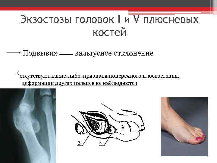 Exostoses of the heads of the I and V metatarsal bones Subluxation valgus deviation
